
Development
Chapter 4 – The Brain and Neuropsychology
The temporal lobe is the side portion of each hemisphere and is located near the temples. It is the primary target for auditory information, so is essential for understanding spoken language. It is involved in advanced visual processing and plays a part in emotional and motivational behaviours.
The occipital lobe is located at the back of the brain and is the main target for visual information.
The frontal lobe is located at the front of the brain and contains the primary motor cortex and the pre-frontal cortex. The frontal lobe is mainly involved in planning of movements, recent memory, and some aspects of emotion such as aggression. It is said to be crucial for planning and controlling thoughts and behaviour.
The parietal lobe is located at the top of the brain between the frontal lobe and occipital lobe. It is responsible for bodily sensations and monitors all the information about eye, head and body positions before passing it on to the brain areas that control movement.
The cerebellum is a large hindbrain structure located below the occipital lobe. It contributes to the control of movement and is important for balance and coordination.
Lateralisation of function
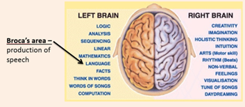
Asymmetrical function: Both sides of the brain are not a mirror image of each other. Each hemisphere controls different functions, or plays a larger or smaller role in a particular behaviour – they are not equal in what they do. E.g.: The right hemisphere controls spatial awareness (ability to negotiate space and navigate our way around our environment)
Each hemisphere controls the opposite side of the body. They are joined by a layer of nerve fibres called the Corpus Callosum through which the two hemispheres can communicate while still working independently.
Gender Differences in Lateralization – There is some evidence that females have a thicker corpus callosum than males
- Females tend to use both sides of their brains for tasks
- Males tend to show dominance for one hemisphere rather than an equal spread
- This means males are more affected by brain damage to one side of their brain, whereas women are less affected by the same brain damage.
- Women are thought to be better at language tasks whereas men are supposed to better at spatial
Strengths –
- Lot of scientific research conducted on differences in males and females allowing it to be highly controlled and reliable
- There is lots of evidence to support it such as Harasty et al. (1997) which suggests the parts of the brain responsible for language are bigger in females and Rilea et al. (2005) found that men were better at some spatial tasks (esp. those that use a lot of activity in the RH)
Weaknesses –
Gender differences aren’t always obvious (e.g.: Rilea et al. (2005) didn’t show men always doing better than women on spatial task.
Some research shows that there is no evidence for differences in brain laterization (e.g.: Sommer et al. (2004) found no strong evidence that females used both hemispheres in language tasks.
Role of the Nervous System
CNS – the brain and spinal cord which relays message from the brain to the rest of the body to instruct it what to do.
PNS – the system of nerves that connect the CNS (mainly the spinal cord) to the skin, muscles and organs in the body.
Neurotransmitters – chemicals found within the nervous system that pass messages from one neuron to another
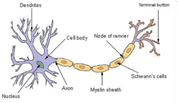
The Neuron
The Synapse
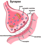
Synaptic Transmission –
- An electrical impulse is triggered inside the cell body of a neuron
- A small impulse is passed along axon (long structure connecting cell body of neuron to terminal button at the end of the cell) towards the end of the nerve
- It arrives at the terminal buttons (end of a neuron) which are filled with small sacs containing neurotransmitters called vesicles
- These contain neurotransmitters
- When the impulse reaches here, the vesicles release their chemical into the synaptic gap
- These chemicals are then grabbed by the receptors (sites on a neuron designed to absorb neurotransmitter molecules) on the next nerve cell, which continues the process to pass the message on.
Neurotransmitters –
| Neurotransmitter | What does it do? | What happens if there is an imbalance? |
| Dopamine | Plays a role in attention and learning | Not enough can make it difficult to concentrate on tasks |
| Serotonin | Plays a role in our mood | Too little can make people feel depressed |
| GABA | Plays a role in calming us down | Not enough causes us to feel stressed |
Neurological Damage
- Damage to the body’s central and peripheral nervous system
- Messages can be interrupted
- Neurons might not be working
- Normal functions of the brain not possible
- Behaviour can be affected
| Visual Agnosia | Prosopagnosia | |
| What is it? | Inability to recognise things that can be seen | Face Blindness |
| Symptoms | Cannot recognize colour, name objects or recognize places | Cannot identify people from their faces or say all faces look the same |
| Brain areas affected | Parietal Lobe | Fusiform Face Area – part of the temporal lobe |
Damage to the Pre-frontal Cortex – The pre-frontal cortex controls aggression and impulses and damage to it causes more impulsive and aggressive behaviour. Adriane Raine et al. (1997) found that murderers have less activity in their pre-frontal cortex compared to a group of people who hadn’t committed murder and used it as an explanation to why some people are more prone to violent behaviour.
Damasio et al. (1994) The Return of Phineas Gage: Clues about the Brain from the Skull of a Famous Patient
Background – In 1848, Phineas Gage was a railway line worker was a calm and well-liked worker who faced an explosion which forced iron rod through his head and turned him into and irresponsible and rude worker who died 12 years later due to epilepsy.
Aim – To investigate the brain damage to Phineas Gage using his skull, in order to determine functions of the frontal lobe
Procedure –
- Gage’s skull was exhumed
- Took pictures and measurements to build a 3D model of skull and iron rod (buried with Gage) – 109 cm long and 3 cm in diameter
- Mapped 20 entry and 16 exit points
- 5 most likely were decided on
- Damage was likely in both left and right hemisphere
- Worst damage in ventromedial region (responsible for sensible decisions)
- Compared to 12 patients with similar damage
Results –
- Extreme damage to the frontal lobe
- Damage to white matter in the left hemisphere (neurons pass their message along their axons which is what white matter consists of) meaning Gage was unable to pass neural messages in this part of the brain
- Damage was worse in the middle of each hemisphere (ventromedial region) than top edges (dorsolateral)
Conclusion – Ventromedial region of the frontal lobe responsible for sensible decisions and emotions. Also responsible for controlling impulses.
Strengths –
- Damasio et al. were able to use modern day technology making the study quite scientific.
- The use of a computer model is more reliable than the reports from 1848.
- We can now make predictions about damage to the frontal lobes.
- We can now treat people who have similar brain damage based on what happened to Gage.
- The study is based on a real-life case of brain damage so is valid.
Weaknesses –
- It is hard to generalise from the study because the brain damage is so unique to Gage.
- The reports of the damage itself are over 150 years old, meaning they may not be reliable.
Sperry (1968) Hemisphere Deconnection and Unity in Conscious Awareness
Background – Patients with severe epilepsy were offered surgery to reduce their fits and due to this had their corpus callosum cut down which led to their brain working into separate halves but no other symptoms were shown
Aim – To find out the cognitive functions that are linked to each hemisphere in the brain.
Procedure –
- 11 patients who had undergone surgery for severe epilepsy.
- Sperry asked patients to fix their eye gaze on the centre of a screen and then projected words or pictures on the right and left side of the visual fields. This way he could be sure that the information only entered either the left or right side of the eye (left eye would pick up image or word on right side of centre). They were then asked to say the word/s or picture/s they had seen. They would be asked to point at an item or picture. They would be shown a variety of objects or pictures and would have to identify what they had seen with either the same hand or opposite hand. They would also have to feel unseen objects and have to describe them and then putting different objects in each hand and have to find them in a large pile.
Results – When words were shown to the LVF (RH) then the patient had trouble saying what they had seen. When words or pictures were shown to RVF (LH), patient had trouble pointing to the object. If objects were felt by the right hand (LH), patient had difficulty naming the object. Patients could identify objects only if they had held it with that hand before, not the opposite.
Conclusions – The left hemisphere was the primary hemisphere for the processing of language (although Sperry showed limited language processing in the right too). The right hemisphere was able to read words (which enabled the patients to recognise objects), make mental associations, process emotional reactions and solve simple arithmetic, and was better than the left hemisphere at spatial skills.
Strengths –
- Sperry gathered a lot of information, which improves the reliability of the study
- The procedure was kept the same for each participant, keeping it standardised and more reliable
- The study tells us a lot about the lateralisation of the brain
Weaknesses –
- It is hard to generalise from the study because the sample size is small and so specific
- We can’t say that split brains represent normal brains
- The study was a lab experiment that lacked ecological validity
- The task lacked validity – our visual fields normally work together, so we would not have these difficulties in real life
Issues and Debates: How has Psychology changed over time?
- William Wundt opened a laboratory in Leipzig, Germany in 1875 to study people’s thoughts
- Hans Berger developed the EEG (electroencephalograph) in 1924 to measure brain activity in a living brain – electrodes are placed on different parts of the scalp to measure general level of activity in different areas of the brain
- Magnetic Resonance Imaging (MRI scans) developed (studies brain using electromagnets)
- Positron Emission Tomography (PET scans) developed (imagery showing amount of energy being used throughout the brain)
- High-powered microscopes allowing psychologists to look at individual synapses are being developed