Transport in Humans
Circulatory System
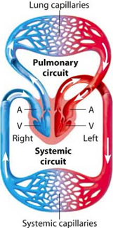
The circulatory system is a system of blood vessels with a pump and valves to ensure one-way flow of blood.
- Single circulation means blood passes through the heart only once i.e. fish
- Double circulation means blood passes through the heart twice i.e. mammals
Double circulation is advantageous because it maintains a higher blood pressure compared to a single circulation system.
Blood Flow Around the Body
All mammals (including humans) have a double circulatory system of blood flow.
- Deoxygenated blood enters the right atrium (RA)
- Deoxygenated blood enters the right ventricle (RV)
- Deoxygenated blood is pumped by the RV to the lungs to become oxygenated
- Oxygenated blood enters the left atrium (LA)
- Oxygenated blood enters the left ventricle (LV)
- Oxygenated blood is pumped by the LV to the rest of the body
- Body cells use the oxygen and cause the blood to become deoxygenated
- Deoxygenated blood returns to the heart and the cycle repeats (step 1)
Heart Structure and Function
Structure of the Heart
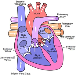
Vena cava → Right atrium → Atrioventricular valve → Right ventricle → Semilunar valve → Pulmonary artery → Lungs →Pulmonary vein → Left atrium → Atrioventricular valve → Left ventricle → Semi-lunar valve → Aorta → Body → Vena cava
Functions of the Different Structures
Atrium – The right and left atrium contracts to pump blood into the right and left ventricles respectively.
Ventricles – The right ventricle contracts to pump blood to the lungs (to become oxygenated). The left ventricle contracts to pump blood to the rest of the body. It has a thicker wall than the right ventricle because it needs to pump blood further, and therefore needs more force.
Atrioventricular valves – The atrioventricular valves separate the atrium and ventricles on both sides of the heart. These valves prevent the backflow of blood, thus ensuring a one-way flow of blood from the atria to the ventricles.
Semilunar valves – Semilunar valves are found within the pulmonary arteries and the aorta. They prevent the backflow of blood and ensures unidirectional blood flow in the arteries.
Pulmonary artery – The pulmonary artery carries blood from the right ventricle to the lungs.
Pulmonary vein – The pulmonary vein carries blood from the lungs to the left atrium.
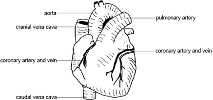
Aorta – The aorta is a large artery which carries blood from the left ventricle to the rest of the body.
Vena cava – The vena cava is a large vein which carries deoxygenated blood from the body back to the heart (right atrium).
Septum – The septum is a thick muscular wall which separates the right and left side of the heart. This separation is important to ensure that oxygenated and deoxygenated blood does not mix.
Activity of the Heart
Physical Activity Increases Heart Rate
Heart rate is the rate at which the heart beats. The most common way to measure heart rate is by measuring the pulse rate. The pulse rate is exactly equal to the heart rate, as the contractions of the heart cause the increases in blood pressure in the arteries that lead to a noticeable pulse.
Physical activity increases the energy demand in muscles such as the arms and legs. With an increased rate of respiration, blood must travel quicker to the muscles to supply them of oxygen/nutrients whilst also removing waste products such as carbon dioxide. The heart rate, therefore, increases to meet these demands.
Electrocardiogram (ECG)
An electrocardiogram is a device which can track heart activity. It can accurately measure pulse rates via the opening and closing of heart valves.
Coronary Heart Disease
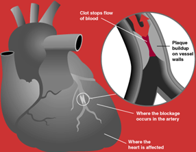
The heart functions as a pump which delivers blood to the rest of the body. However, the heart muscles themselves also need a blood supply because they too, are respiring muscles. The coronary artery is the very important artery which provides the heart muscles with blood.
Coronary heart disease is when the coronary artery becomes blocked, leading to blood (and oxygen) starvation in the heart muscles. This leads to a heart attack.
Causes – Blockage of the coronary artery begins by the narrowing of the artery due to cholesterol build up on the inner walls. Total blockage can occur when a blood clot gets ‘stuck’ in these narrow arteries.
Risk factors – Certain factors that increase the risk of an individual developing coronary heart disease:
- Poor diet
- Stress
- Smoking
- Genetics
- Age
- Gender
Treatment with medications – Blood thinning medications are used to reduce the chances of a blood clot forming
Treatment with surgery –
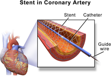
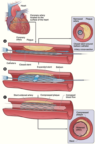
- Stents are a tube-shaped device which is placed inside the coronary arteries to physically hold it open
Angioplasty is a stent with a balloon which can be inflated once the stent is inserted to even further increase the diameter of the artery
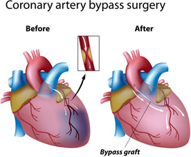
Bypass is the process of making a separate ‘new’ artery to allow for an alternative blood path that the heart can use to receive blood (rather than relying only on the coronary artery)
Blood Vessels
Blood vessels are tubular structures carrying blood through the tissues and organs. Starting from the heart, the pathway of blood is as follows:
[HEART] → Artery → Arteriole → Capillary → Venule → Vein → [HEART]
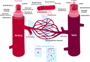
The artery branches out into arterioles which branch further into capillaries which then join to form venules which join further to become the vein.
Arteries – They take blood away from the heart. They have several important structural features:
- Thick muscular walls to withstand blood being carried at high pressures
- Narrow lumen which expands as blood pulsates through to maintain blood pressure
- Valves absent since high blood pressures prevent back flow
Arterioles – They are smaller branches of an artery. They eventually branch further to form capillaries.
- Arterioles have muscular/ elastic walls that can constrict & dilate in order to regulate blood flow.
Capillaries – They are fine branching blood vessels that form a network between the arterioles and venule.
They allow for the nutrient & waste exchange between the blood and the tissues of the body. The features of capillaries are as follows:
- Walls are one cell thick to allow for quick diffusion of nutrients/wastes
- Lumen has a diameter of just one RBC to allow blood cells to pass closely to the walls for faster diffusion rates
- Valves are absent since the narrow capillary lumen ensures unidirectional blood flow
Venules – They are small vessels formed from the joining of the capillaries. Venules combine to establish a vein.
Veins – They take the blood towards the heart. Their structural features are as follows:
- Thin walls with little muscle & elastic fibres (thick muscles not required since blood is carried at low pressure)
- Large lumen to reduce blood flow resistance
- Valves present to prevent blood back flow
Shunt vessels – Blood vessels that connect blood directly from the arterioles to the venules. This allows for an alternative route for blood flow (i.e. blood bypasses the capillaries).
Like arterioles, shunt vessels have walls that can construct & dilate in order to regulate blood flow.
Tissue Fluid
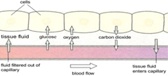
Tissue fluid is the fluid which bathes most body tissues. The fluid is the mode of nutrient & waste exchange between the blood and respiring tissues.
For example:
- Waste products (such as carbon dioxide) from cells diffuse into the tissue fluid first before diffusing in the blood
- Nutrients (such as glucose) diffuse into the tissue fluid first before diffusing into the cells
Tissue fluid is produced by leakage of certain substances from blood capillaries, and drained out by the lymphatic vessels of the lymphatic system.
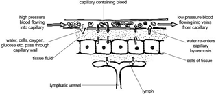
NOTE: Details of tissue fluid production and drainage are NOT required. Just understand that blood capillaries LEAK tissue fluid and the tissue fluid is DRAINED by lymphatic vessels. The tissue fluid inside lymph vessels is called LYMPH.
The lymphatic system is composed of lymphatic vessels which carry “lymph” and lymph nodes which produce lymphocytes for immunity.
Blood
Blood is a mixture of several components such as:
- Red blood cells (RBCs)
- White blood cells (WBCs)
- Platelets
- Plasma
Plasma – Blood plasma makes up about 50% of the blood. It is a yellowish liquid that carries the other blood components such as RBCs/WBCs/platelets.
Red blood cells – RBCs contain haemoglobin which binds to oxygen for transportation around the body.
White blood cells – WBCs are a part of the immune system that helps to destroy foreign organisms such as bacteria.
There are two types of white blood cells that you need to be aware of:
- Phagocytes are types of WBCs which engulf and digest pathogens via phagocytosis
- Lymphocytes are WBCs which produce antibodies
Platelets – Substances that form blood clots which is a protective mechanism to prevent blood loss during an injury.
At the site of damage, platelets immediately stick together and release chemical signals which attract other nearby cells and clump them together.
A series of chemical reactions take place. Fibrinogen is converted into fibrin and this forms a thread which traps RBCs to establish a thick clot. The clot seals off the site of damage.