Cells
Cell Structure and Organization
First of all, all organisms are made of cells. They are like the lego blocks of life. The syllabus wants you to know how to draw a basic animal and plant cell, label its structures and also explain the functions of each of the structures too.
At a very basic level, please refer to the diagram below. The plant cell has everything that an animal cell has, plus some added structures which are written in green text. The functions of each of these structures will be discussed further down the page.
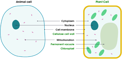
All cells have a cell membrane (thin layer of protein and fat) which is what allows or disallows certain things entering and exiting the cell (and is therefore partially permeable).
The nucleus contains genetic information (DNA) about the right sorts of proteins to be made in the cell. Chromosomes made of DNA are very long but so thin that they cannot easily be seen even using the electron microscope unless the cell is dividing, because they become short and thick, and can be seen with a good light microscope.
The cytoplasm is a jelly-like substance (about 70% water) containing various dissolved substances (esp. proteins) where many metabolic reactions take place.
The mitochondrion is the “powerhouse’ of the cell and the reason for this name is due to the fact that respiration occurs here and cells that use a lot of energy have a lot of mitochondria (e.g.: muscle cells; sperm cells – which need energy to swim to the egg; and neurons/ nerve cells – which need energy to transmit impulses).
Plants have some extra structures such as cell walls made of cellulose (to support the cell and stop it from bursting in case of the cell swelling with water). Cellulose is a polysaccharide and forms crisscross fibres with large spaces in between due to which even large molecules can go through and the cell wall is, thus, fully permeable.
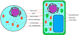
They also have chloroplasts containing chlorophyll which absorb energy from sunlight which is later used for photosynthesis. Chloroplasts may also contain starch grains – animal cells have tiny granules of glycogen. Glycogen is a reserve fuel (similar to starch but animal cells do not store starch). When required, it can be broken down to glucose, to be used as a fuel by the mitochondria in the liver cell, or transported in the blood to other cells that need it and is found in the cytoplasm.
Plants also have a permanent vacuole (space in the cell) containing cell sap – solution of sugar and other substances – and keeps the cell in shape, whereas animal cells have small temporary ones called vesicles, which may contain food or water. The Rough Endoplasmic Reticulum (RER) is a set of tubular membranes near the nucleus which have ribosomes studded onto them, and the ribosomes are then used for protein synthesis (joining of amino acids in long chains according to instructions carried on DNA in the cell’s nucleus). These ribosomes can either be found on the RER (as mentioned before) but they can be found freely in the cytoplasm as well.
Structure & Function Summary
- Cell Membrane – Selective control of what goes in and out of the cell
- Nucleus – Carries genetic material (DNA)
- Cytoplasm – Jelly like substance in which chemical reactions take place
- Cell Wall – Structural support for the cell
- Chloroplast – Site of photosynthesis
- Vacuole – Storage of nutrients
- Rough Endoplasmic Reticulum – Studded with ribosomes
- Ribosomes – Site of protein synthesis
- Mitochondria – Site of aerobic respiration (cells with high metabolism rates will need lots of these to offer sufficient energy)
Levels of Organization
There are levels of organization that you need to be aware of. As we discussed before, the smallest unit of a living thing is a cell. So that’s a good place to start. A group of cells is called a tissue, a group of tissues is called an organ, and a group of organs is then called an organ system.
Take a look here:
- Cell – Smallest structural and functional unit of an organism
- Tissue – Group of cells with similar structures working together to perform a shared function
- Organ – Structure made up of a group of tissues, working together to perform specific functions
- Organ system – Group of organs with related functions, working together to perform body functions
Now some cells have structures that help them with their particular function such as:
- Ciliated cells – move mucus upward
- Root hair cells – absorb water and mineral salts
- Xylem vessels – transport water and mineral salts; help in support
- Palisade mesophyll cells – photosynthesis
- Nerve cells – transmit information in the form of electrical impulses
- Red blood cells – transport oxygen
- Sperm and egg cells – fuse together to produce a zygote
Each of the things above will naturally be covered in more detail in other topics in the syllabus and therefore will not be covered here.
Size of Specimen
In the lab, a lot of biology is done under a microscope. For example, we can’t examine the cells of human tissue with our naked eyes, right? Therefore, the purpose of the microscope is to magnify our specimen so that it appears bigger for us to be able to see. A light microscope shines light through the piece of animal or plant you are looking at and uses glass lenses to magnify and focus the image. A very good light microscope can magnify about 1500 times. A photomicrograph is a picture made using a light microscope. To see even smaller things inside a cell, an electron microscope is used. This uses a beam of electrons instead of light and can magnify up to 500,000 times. This means that a lot more detail can be seen inside a cell. We can see many structures more clearly, and also some structures that could not be seen at all with a light microscope in electron micrographs (pictures taken from an electron microscope).