
Cell Structure
Microscopy
● Light microscope-uses light as a source of radiation
● First used in beginning of 17th century
● Early 19th century, quality of lenses improved creating dramatic images
● Cytology-study of cells
● By 1900, all cell structures and organelles except lysosomes had been discovered
Units of Measurement in Cell Studies
● International System of Units (SI) only accepted scientific measurement system
● Basic unit of measurement – meter (m)
●
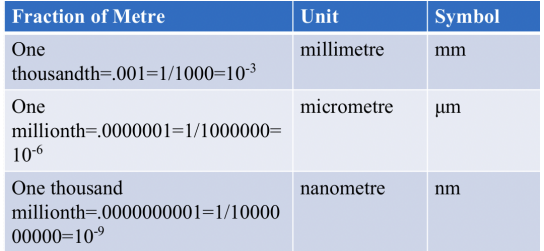
SI Units of Measurement
● 1 micrometre (μm) is a thousandth of a millimetre (mm)
● 1 nanometre (nm) is a thousandth of a micrometre (um)
● Cell sizes range from 5 μm to 40 μm
● Smallest structure visible with only the human eye is about 50-100 μm.
● Smallest organelle, ribosomes, are only about 20 μm in diameter.
Sizes of Biological Structures
● Smallest object visible with eye only – 50-100 μm, about diameter of sharp end of a pin
● Smallest object visible with a light microscope – 0.2 μm or 200 nm, about the average size of a bacterium
● Smallest object visible with an electron microscope – 0.5 nm, ribosome, cell membrane
● Invisible – diameter of a hydrogen atom (smallest atom) 0.04 nm
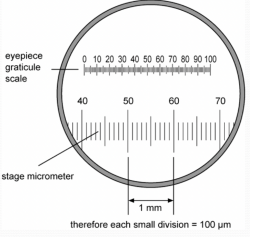
Measuring Cells
● Using Stage Micrometer this cell measures 1 μm, marked in .1 μm and 1 μm divisions
● 1 cm = 10 mm = 1000 micrometers (μm)
Calibrating Eyepiece Graticule
● Using an eyepiece graticule with arbitrary scale – must calibrate to stage micrometer to
determine actual measurement.
● Count number of divisions on EG to equal to 10 μm on stage micrometer
Calibrating Eyepiece Graticule
● On lowest power, count the number of divisions on eyepiece graticule equal to 10 μm on the stage micrometer to calculate length that one eyepiece division is equal to.
● For example, if 43 divisions are equal to 10 μm, then each division is equal to 0.233 μm at low power.
● Repeat for medium and high power objectives.
Magnification versus Resolution
● Magnification-number of times larger an image is compared with the real size of an object.
● Magnification = measured size of magnified image / actual size of specimen
Resolution
● Ability to distinguish between two separate points
● If two points can’t be resolved, they’ll be seen as one point
● Maximum resolution of light microscope is 200 nm
● The limit of resolution is one half the wavelength of the radiation used to view the specimen
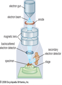
Electron Microscopy
● Free electrons behave like electromagnetic
radiation – they have a very short wavelength
● Suitable form of radiation for microscopy due
to:
- Extremely short wavelengths (think
X-rays) - Negatively charged, focus using
electromagnets
Types of Electron Microscopes
● Transmission - Beam passes through specimen
- Only electrons transmitted (through) specimen seen
- Advantage-can view inside of cells/structures
- Can view thin specimens
● Scanning - Scans surfaces of specimens
- Only reflected beam is observed
- Advantage – surface structures seen with great depth of field
- Disadvantage-resolution not as good as TEM
Overall Difficulties with Electron Microscopy
● Must take place in a vacuum. Air molecules would cause electrons to
scatter
● Water boils at room temperature in a vacuum, so all specimens must be
dehydrated
● Only dead material can be examined with EM