Cell Structure
Cytology – Study of cells
Animal Cell Structure:
Organelles:
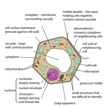
Nucleus:
- Largest organelle and is stained easily.
- Surrounded by two membranes – nuclear envelope.
- On the nuclear envelope are small nuclear pores which allow exchange between cytoplasm and nucleus. Example: Protein
- Chromosomes (contain genetic material) are only visible during cell division.
- Nucleolus (composed of DNA and RNA) inside the nucleus makes ribosomes from its own DNA.
Endoplasmic reticulum (ER): Network of flattened sacs running through the cytoplasm of eukaryotic cells. Around the nucleus connecting to the nuclear membrane.
Two types:
- Rough ER: Covered with many ribosomes.
- Smooth ER: Lacks ribosomes and makes lipids, steroids such as cholesterol. Reproductive hormones such as oestrogen and testosterone are also made here.
Ribosomes (25μm): Have Large and small subunits.
- 70s in prokaryotes, 80s in Eukaryotes.
- Not membrane bound.
Golgi Body/complex/apparatus:
- Collects, processes and sorts molecules.
- Transports them in Golgi vesicles to other parts of cells.
- Used to make lysosomes.
- Network of membrane bound vesicles.
Golgi Vesicles: Carry their contents to other parts of the cell, to the cell surface membrane for secretion.
Lysosomes: Spherical sacs surrounded by a single membrane (0.1-0.5 μm)
- Produces hydrolytic enzymes which breaks down old cells or organelles, for example, Mammary glands.
- In WBC, lysosomes digest bacteria.
Mitochondria (1μm): Rod /sausage/cylindrical shaped.
- Provides energy for respiration, inner membrane is folded to increase surface area.
- Most numerous organelles seen with a light microscope.
- Carry out aerobic respiration.
- Has folds in the inner membrane called cristae which are found on which stalked particles of ATP synthase.
Microtubules:
- Long, rigid, hollow tubules found in cytoplasm. They are part of the cytoskeleton.
- Made from tubulin (protein), alpha tubulin and beta tubulin.
Centrioles:
- Hollow cylinder about 500nm long.
- Formed from a ring of short microtubules.
- Has 9 triplets of microtubules.
Cilia: Whip like structures projecting from the surface of many animal cells. They beat and allow the movement of cells across a fluid.
- They are composed of 600 different polypeptides.
- Has a basal Body, cilium, cell surface membrane.
- Have two central microtubules and a ring of nine microtubule doublets.
Microvilli: Small finger-like extensions of a cell that increase the surface area of the cell for more efficient absorption or secretion.
Vacuoles:
- Sac like structure surrounded by a single membrane.
- Plant cells have a large permanent vacuole, animal cells have temporary vacuoles.
- Plant vacuole is surrounded by tonoplast which controls exchange between vacuole and cytoplasm.
- Fluid is a solution of pigments, enzymes, sugars etc.
- Helps to regulate osmotic properties.
Secondary Metabolites:
- Anthocyanins are pigments that are responsible for most of red, purple, pink and blue colours of fruits and flowers.
Chloroplasts:
- Organelles in plant cells responsible for photosynthesis.
- Bounded by an envelope.
- Grana inside consist of chlorophyll.
- Tend to have an elongated shape. (3-10 μm).
- Membrane system consists of fluid filled sacs and thylakoids which spread out like sheets in three dimensions.
- Contains 70s ribosomes and circular DNA.
- Have Thylakoids: Flattened membrane bound fluid filled sac which is the site of light dependent reactions of photosynthesis in a chloroplast.
Cell Walls:
- Relatively rigid. Consists of parallel fibres of the polysaccharides such as pectins and hemicelluloses.
- Have high tensile strength and are inelastic.
- Xylem vessel elements are example of sclerenchyma.
Functions:
- Mechanical strength and support for individual cells and the plant as a whole. Turgid tissues.
- System of interconnected cell walls in a plant is called the apoplast , it is the major transport route for water, inorganic ions and other materials.
- Epidermal cells often have a waterproof layer of waxy cutin, the cuticle, on their outer walls. This helps reduce water loss by evaporation.
Prokaryotes and Eukaryotes:
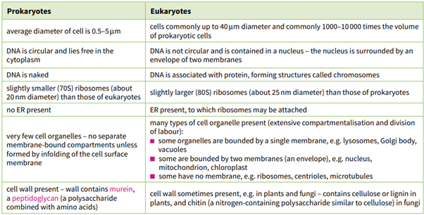
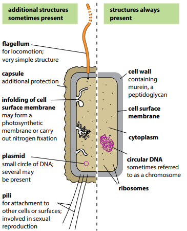
Bacteria:
- Cell walls contain peptidoglycan.
- Has cell surface membrane.
- Cytoplasm does not contain any double membrane bound organelles.
- DNA molecule in bacteria is circular, found in the nucleoid which also has small amounts of RNA.
- Bacterial ribosomes are 70s.
- Bacterial flagellum is a simple hollow cylinder made of identical protein molecules.
- Some bacteria are surrounded by a cell wall which makes a slimy layer.
- A plasmid is a small circle of DNA separate from the main DNA of the cell. Plasmids can copy themselves independently of the chromosomal DNA and can spread rapidly from one bacterium to another.
Pili are fine protein rods.
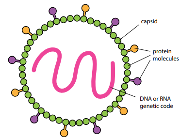
Virus:
- Very small (20-300nm) infectious particle which can replicate only inside living cells; it consists of a molecule of DNA and RNA surrounded by a protein coat.
- Protective coat of protein is called capsid.
Microscopes:
- Electron microscopes:
- Only dead material or non-living can be examined.
- Electron bean is used so it must be in a vacuum, because if the electrons collided with air molecules the picture will not be sharp.
- Water boils at room temperature in a vacuum so only dehydrated, dead organisms can be used.
Two types:
- TEM: Transmission electron microscope, internal structures of cells and organisms can be seen.
- SEM: Scanning electron microscope, scans the surfaces of structures and only the reflected beam is observed.
Resolution: Ability to distinguish between two objects very close together, the higher the resolution of an image, the greater the detail that can be seen.
Magnification = Image size/ Actual Size.
Note: All images and some text have been adapted from Cambridges AS and A level Biology Coursebook.