
Cell membrane and transport
Cell membrane:
- Is partially permeable
- Thin
- Function example: enable cells to receive hormone messages.
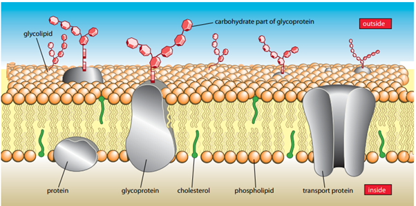
Made of phospholipids, proteins, carbohydrate chains. - Regulates exchange of substances.
Structure:
Phospholipids:
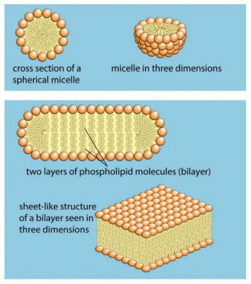
- Hydrophobic tail(non-polar), Hydrophilic head (polar).
- Phospholipids can form ball like structures called micelles or can form a bilayer.
- Bilayer is 7nm wide.
- Water soluble molecules such as sugars cannot enter the cell, nor can they leak out.
Fluid Mosaic model:
- “Fluid” because both phospholipids and proteins can move around by diffusion. Phospholipid molecules move sideways.
- “Mosaic” because it is the pattern produced by protein and other molecules on the surface of the membrane.
- Nonpolar interior of the membrane.
- Some tails are unsaturated and saturated. Greater unsaturated tails, more fluidity. Longer the tail, less the fluidity.
- When temperature decreases, fluidity also decreases.
Cholesterol:
- Have hydrophobic tail and hydrophilic tail.
- Less common in plant cell membranes and absent from prokaryotes.
- Provides mechanical stability.
- Hydrophobic regions prevent leakage of ions in nerve cells.
- Prevents phospholipids packing close together at low temperatures and maintains stability and prevents phospholipids from moving further away at higher temperatures.
Glycolipids and glycoproteins:
- Combination of molecules.
- Carbohydrates chains help glycoproteins and glycolipids to act as “receptor molecules”
- Signalling receptors – part of a signalling system that coordinates cell activities.
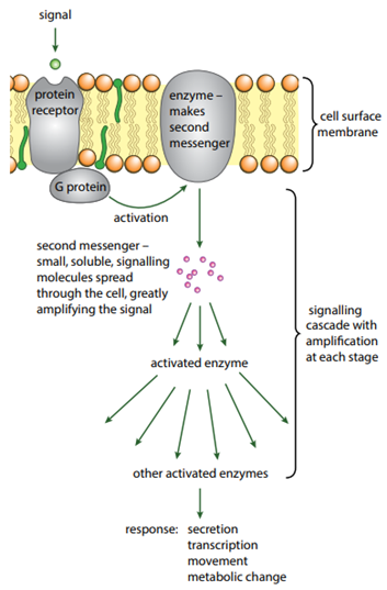
Cell Signalling: The molecular mechanism by which cells communicate with each other and detect and respond to external stimuli.
- A stimulus will cause the secretion of ligand, example, glucagon. Glucagon is secreted as a response to lower blood sugar levels.
- Ligand is transported to “target cells”.
- Ligand binds to cell surface receptors on the target cells.
Ligand: Biological molecules which binds specifically to another molecule, Example: cell surface membrane receptor.
Transduction: Occurs during cell signalling and is the process of converting a signal from one method of transmission to another.
G protein:
Movement of substances into and out of the cell:
Diffusion: Net movement of molecules from a region of higher concentration to a region of lower concentration down a concentration gradient as a result of the random movement of particles.
Factors affecting diffusion:
- Steepness of concentration gradient: Steeper the concentration gradient of a substance, faster the rate of diffusion.
- Temperature: Molecules diffuse faster at higher temperatures due to increased kinetic energy.
- Nature of molecules or ions: smaller molecules diffuse faster and non-polar molecules such as glycerol diffuse more easily through cell membrane because they are soluble in non-polar phospholipid tails.
- Surface area: Greater the surface area, more ions dissolve through the membrane.
Facilitated diffusion: Diffusion of a substance through a transport protein in a cell membrane, the protein provides hydrophilic areas that allow the molecule or ion to pass through the membrane which would otherwise be less permeable.
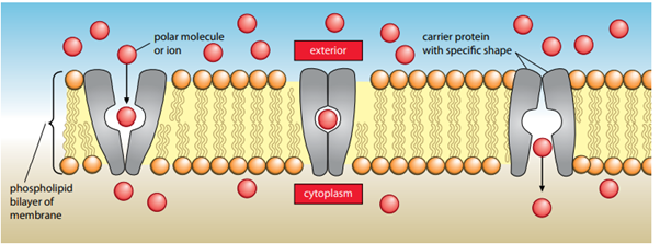
Takes place through:
- Channel protein: A membrane protein of fixed shape which has a water filled pore through which selected hydrophilic ions or molecules can pass by facilitating diffusion or active transport.
- Carrier protein: Change’s shape to allow specific ions or molecules to pass through the cell membrane by active transport or facilitated diffusion.
Osmosis: The net diffusion of water molecules from a region of water potential to a region of lower water potential down the concentration gradient, through a partially permeable membrane.
Water potential: A measure of the tendency of water to move from one lace to another; water moves from a solution with higher water potential to a region of lower water potential.
- Water potential decreases when more solute is added.
- Water potential increases with increased pressure.
- Symbol for water potential; psi Ψ.
- In plant cells, Water potential is Ψ= Ψs+ Ψp
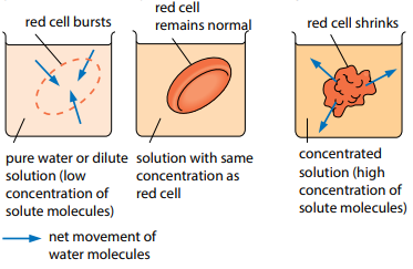
Animal cells will burst when placed in dilute solution due to lack of cell wall. As water molecules move from the region of higher concentration outside the cell into the region of lower concentration in the cell, the increased pressure causes the cell to burst.
Similarly, an animal cell will shrink when placed in concentrated solution as water molecules are drawn out.
- Plant cells are surrounded by cell walls which have high tensile strength and are strong and rigid.
- When plant cells are placed in concentrated solution, the protoplast shrinks as water is lost and the vacuole becomes flaccid. Cell is plasmolysed.
- When plant cells are placed in a dilute solution, the protoplast expands, and the vacuole becomes turgid.
Plasmolysis: The loss of water from a plant or prokaryote cell to the point where the protoplast shrinks away from the cell wall.
Incipient Plasmolysis: Point at which plasmolysis is about to occur when a plant cell or a prokaryote cell is losing water; at this point the protoplast is exerting no pressure on the cell wall.
Active Transport: Movement of molecules or ions through transport proteins from a region of lower concentration against their concentration gradient using energy from ATP.
- Important in reabsorption in the kidneys where useful molecules and ions are reabsorbed into blood after filtration in kidney tubules.
Carrier protein example:
- Sodium potassium pump – A membrane protein that moves sodium ions out of the cell and potassium ions into the cell using ATP.
Endocytosis – Bulk movement of liquids (pinocytosis) or solids (phagocytosis) into a cell by the infolding of the cell surface membrane to form vesicles containing the substance. Requires energy
Exocytosis: Bulk movement of liquids or solids out of the cell by the fusion of vesicles containing the substance with the cell surface membrane. Requires energy.
Phagocytosis takes place in phagocytes.
Phagocytes: Cell which ingests and destroys pathogens or damaged body cells by phagocytosis. Example WBC.
Note: All images and some text have been adapted from Cambridges AS and A level Biology Coursebook